
The NeuronThe brain contains two types of cells: nerve cells and glial cells. Nerve cells (also known as neurons) carry nerve signals to and from the brain and aid in thought, memory, emotion, speech, and muscle movement. In contrast, glial cells are the supporting cells of the brain and provide nutrients and insulation for neurons. Since alcohol will primarily affect the transmission of the signal from one neuron to the next, we will not go into details about the glial cells.
In the body, there are as many as 10,000 specific types of neurons used to convey different types of signals. The image below illustrates a few of them (Stufflebeam, 2008). 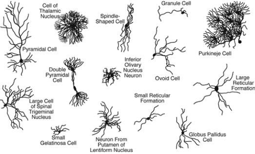
|
A typical neuron is comprised of the following parts: 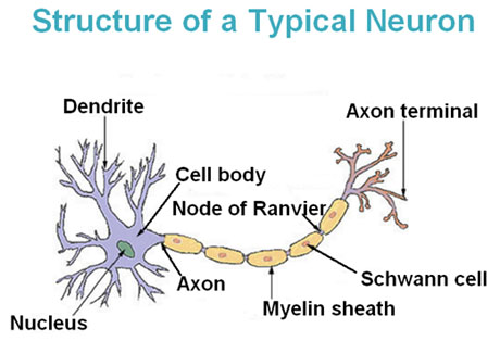
Signals from the brain or other adjacent neurons come in through the dendrites. These signals get processed in the cell body and then flow through the axon to the axon terminal. In the case of a drug, such as alcohol, the axon terminal is an important structure to note. It is located at the end of the axon and contains neurotransmitters. Neurotransmitters are responsible for the chemical signal that tells one neuron to conduct a signal. They allow the projection of the signal to the next dendrite or to its destination in the body.
|
Action Potentials
Communication between neurons is of utmost importance for the body to carryout any action. As such, neurons have a unique way of signaling each other. This signaling is accomplished through action potentials. Action potentials are much like electrical signals in that they travel along the axon, like electricity down a wire. The membrane potential of a neuron is maintained by ion concentrations inside and outside the cell. Ion channels open and close to allow ions to pass in and out of the cell to maintain a constant resting membrane potential of -70 mV. Since the membrane is negative, this state is called polarized. When the neuron get stimulated, the sodium channels open and a large influx of sodium ions go into the cell. At this stage, the cell becomes depolarized. This influx of positive charge causes the cell's potential to become positive. The spike in charge is called an action potential. Once the potential goes up to roughly +30mV the sodium channels close (Stufflebeam, 2008). Then, the potassium channels open and potassium ions move outside the cell. As potassium ions leave the cell, the membrane potential of the cell starts to repolarize. Consequently, the changes in polarity of the cell cause adjacent ion channels along the cell membrane to open and close. The animation below shows a graph of the membrane potential as the ion channels open and close. This process continues through the axon and is how a signal propagates through a cell.

Animation from (Stufflebeam, 2008)
NEUROTRANSMISSION
So, how do cells know what kind of signals to send?
The answer to this question are the different types of neurotransmitters that are released from the axon terminals of one neuron and received by the dendrites of an adjacent neuron. Between neurons there is a small gap call the synaptic cleft. It separates the presynaptic neuron from the postsynaptic neuron. Generally, there are two types of neurotransmitters: excitatory and inhibitory. The brain communicates to the body via different neurotransmitters that affect mood, sleep, concentration, digestion, heart beat, and many more functions of the body. As the concentrations of excitatory and inhibitory neurons change, such as with the introduction of substances like alcohol, they can have a broad range of affects on mental cognition.
How do they work?
The conduction of the action potential prompts the release of a vesicle with a neurotransmitter inside to release into the synaptic cleft from the presynaptic neuron. Ion channels on the postsynaptic neuron only open when a specific neurotransmitter binds to the receptor site on the channel. Once the neurotransmitter binds to the channel, it opens and allows for the influx of ions into the postsynaptic neuron, continuing the conduction of an action potential. After a certain time period, the neurotransmitters are taken up by the presynaptic nerve using transporters in a process called re-uptake. The re-uptake of the neurotransmitters allow for the neurotransmitters to be recycled. The animation below gives a summary of neurotransmission.
Animation from (Stufflebeam, 2008)
Summary (Stufflebeam, 2008)
1) Action potential arrives at the axon terminal.
2) Release neurotransmitter from a vesicle.
3) Neurotransmitter binds to receptor site on ion channel.
4) Ions cross the membrane through open channels, which produces a synaptic potential in the postsynaptic neuron.
5) Neurotransmitter is removed from the cleft.
Types of Neurotransmitters
There are many types of neurotransmitters within the human body and they each have different effects depending on the type. As noted before, all neurotransmitters can be generalized into two categories: excitatory and inhibitory. This description stems from the type of effect the chemical neurotransmitter has on the body, or in particular, the receptor to which the neurotransmitter binds. Because there is an abundant number of neurotransmitters, the table below shows only the most common neurotranmitters and their effects.
| Neurotransmitters | Effects |
|---|---|
| Acetylcholine (Ach) | Distributes widely throughout the central nervous system, where it is involved in arousal, attention, memory, motivation, and movement. Involved in muscle action through presence in neuromuscular junctions (specialized type of synapse where neurons connect to muscle cells). Degeneration of neurons that produce ACh have been linked to Alzheimer's disease. Too much can lead to spasms and tremors; too little can lead to paralysis or torpor. |
| Dopamine | Involved in a wide variety of behaviors and emotions, including pleasure. Implicated in schizophrenia and Parkinson's disease. |
| Serotonin | Involved in the regulation of sleep, dreaming, mood, eating, pain, and aggressive behavior. Implicated in depression. |
| Norepinephrine | Affects arousal, wakefulness, learning, memory, and mood. |
| Endorphins | Involved in the inhibition of pain. Released during strenuous exercise. Also, responsible for runner's high. |
| Glutamate | Involved in long term memory and perception of pain. |
| GABA (Gamma aminobutyric acid) | A largely inhibitory neurotransmitter distributed widely throughout the central nervous system. Implicated in sleep and eating disorders. Low levels of GABA have also been linked to extreme anxiety. |
| Glycene | Principally responsible for inhibition in the spinal cord and lower brain centers. |
Table Citation: (Morris,2000)
